Clone of Upcoming Arrival of Micro-CT
Novel imaging device for studying human fossils
Micro-Computed Tomography (Micro-CT)
Micro-computed tomography (Micro-CT) is a high resolution imaging technology that is increasingly used in the study of fossils, bones, and other irreplaceable archaeological artifacts. Micro-CT is a non-destructive X-ray imaging system, which is a fundamental tool in anthropological research, allowing us to create an exact virtual model of the object and reconstruct the internal and surface structures for further visualization and analysis. Micro-CT is capable to obtain high-quality morphological information preserving the fragile and unique human fossils for future generations.
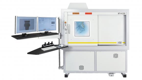
The Imaging laboratory at the Shmunis Family Anthropology Institute operates a Nikon XT H 225 ST X-Ray Micro-Computed Tomography (Micro-CT) system.
Technical specifications
- XT H 225 ST Reflection + Transmission Dual Target system with MRA, PE 1621 EHS, carbon fibre window and motorised FID
The system has two interchangeable sources: the Ultrafocus (reflection) target operates between 20 and 225 kV. The Nanofocus transmission target operates between 20-180 kV to give a micron-sized spot leading to sub-micron feature recognition. 5 axis manipulator with a maximum travel range of 300 mm in X, 350 mm in Y and 750 mm in Z, allows handling of large sample sizes up to 50 kg weight.

